Real-Time Biomedical Computing
Background
Biomedical applications receive increasing attention over the last few years. Driven by the advances in algorithms and computer architecture, ever more realistic simulation of biomedical problems is possible at interactive rates. Especially, ongoing demands are made to accurately predict the behaviour of biomechanical materials due to external forces. Such approaches have a huge application area in all kind of medical simulators. The goal of this project is to develop prototypes for biomechanical material simulation that are ready to be applied in practical applications. Therefore, we are working in close cooperations with medical residents from the Klinikum rechts der Isar der Technischen Universität München.
Visual Computing
What is Visual Computing? In our understanding, Visual computing means to combine computing with visualization. While computing is an extensive term, in our research we focus on numerical simulation, which is actually a very compute-intensive application. Then, Visual Computing means to tightly couple numerical simulation environments with visualization techniques. This opens the possibility to interactively change simulation parameter, thereby directly leading to Computational Steering approaches.
Our research goals are the following:
- Applying advanced methodic and algorithmic techniques to speed-up numerical simulation.
- Developing interactive, yet high-quality, visualization modules by efficiently employing graphics hardware.
We have developed a multigrid framework for the simulation of deformable bodies, which is capable of physical accurate simulation at interactive rates. The framework is based on an implicit finite element formulation, and it incorporates different measures of the mechanical strain of the material ranging from linearized Cauchy strain to non-linear Green strain. The core of the framework is a geometric multigrid solver, which operates on a hierarchy of unstructured grids. Due to the non-linearity of the underlying equations, the multigrid solver has been extended to efficiently handle dynamic matrices - this are matrices which have to be updated in every simulation frame, but typically maintain their sparsity structure. We have developed an acceleration structure that allows to efficiently update the multigrid solver once the underlying matrix has been changed.
Computational Steering for Orthopedics
This research projected, which is financed by the International Graduate School of Science and Engineering, is in close cooperation with our partners from the Chair for Computation in Engineering and the Clinic for Orthopedic Surgery, Sport Orthopedics and Trauma Surgery. The project focuses on predicting and monitoring in-vivo bone strength, which is important in a number of clinical applications ranging from fracture fixation to endoprothesis for joint replacement. To avoid adaptive remodeling with cortical thinning and increased porosity of the bone due to stress shielding, in a pre-operative planning process the optimal implant design, size, and position has to be determined. This process involves interactive implant selection and positioning within the patient-specific bone as well as simulation and visualization of the stress within the bone and implant due to exerting forces.
We employ a multigrid technique to speed up a finite element simulation of the bone based on hexahedral elements. The highly efficient solver allows us to compute the stress distribution in a patient-specific bone at the CT resolution within roughly one minute. From each CT voxel, the simulation determines the respective finite element's elastic modulus. To visualize the resulting 3D stress volume, we apply a volume ray-caster which determines the von-Mises norm for each stress tensor.
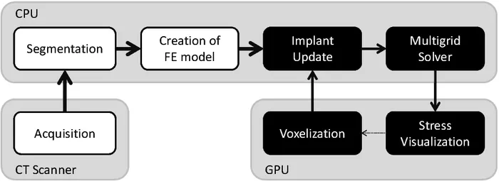
One of the main challenges in this project is the efficient modeling of the implant, since the shape and position of the implant are changed by the surgeon. Therefore, instead of precisely computing the contact between the implant and the holed bone, the simulation is based on a voxelization of the implant with respect to the bone's CT grid. The voxelization of such an implant can be computed efficiently on the GPU within less than 1ms. It is then used to adjust the material parameters of the respective finite elements in the simulation grid, thereby achieving the same behavior as a bonding contact between bone and implant. The employed multigrid finite element framework supports such update operations very well.


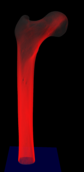
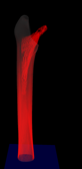
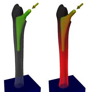
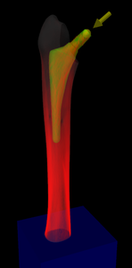
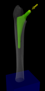
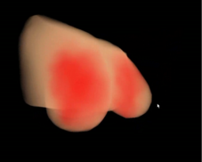
Visualization of the stress distribution inside the bone at different FE model resolutions, with and without the implant.
The resulting prototype allows the surgeon to interactively select varying implants and positions them arbitrarily, thereby supporting the surgeon in the pre-operative planning. Some of the features are demonstrated in a captured interaction with the system.
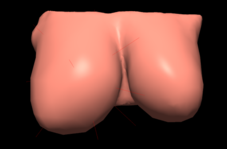
Sinus 2 - Advances in Breast Surgery
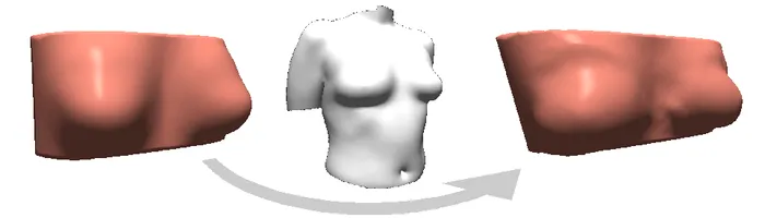
The goal of this research project is to prototype a simulator that supports in plastic and reconstructive surgery of the female breast. We cooperate with the Klinik und Poliklinik für Plastische Chirurgie und Handchirurgie, CADFEM and Steinbichler Optotechnik. Our simulation environment is based on a so-called breast template, which is a finite-element model of the breast, where different stiffness values are assigned to each tetrahedral element based on the type of the underlying tissue (muscle, skin, adipose tissue). The model is adapted to a specific patient by deforming it according to the patient's surface Laser scan. The adapted tetrahedral model is then used in all further steps. To support for patient specific volumetric information that cannot be obtained from the surface scans, the surgeon can choice templates according to further a-priori information available, such as portion of muscle or adipose tissue.
The simulation environment then allows to simulate deformations of the female breast due to external forces such as gravity. Furthermore, virtual implants can be incorporated to predict the post-operative appearance of the patient-specific breast. Implants are modelled through artificial force fields in the interior that can be positioned by the surgeon, and that are carefully adapted to model the kind of implant (anatomic vs. round) and the implant volume selected by the surgeon. By combining the simulation engine with advanced techniques for volume rendering of unstructured tetrahedral meshes, the surgeon is supported in ensuring that tissue close to the implant is not stressed too much.
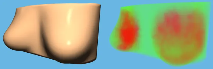
Moreover, the surgeon can potentially minimize post-operative complications (e.g., swelling, bleeding) by optimizing the surgical approach regarding implant parameters, implant position and operative access taking the material properties of the different breast soft tissue types into account. On the other hand, the patient and the surgeon can get an impression of the post-operative appearance regarding volume and shape of the breast. A prospective pre-operative surgical planning and quality assurance of the surgical outcome is possible.

Publications
- A Hexahedral Multigrid Approach for Simulating Cuts in Deformable Objects
- A Real-Time Multigrid Finite Hexahedra Method for Elasticity Simulation using CUDA
- Interactive Deformations with Multigrid Skeletal Constraints
- A Streaming Approach for Sparse Matrix Products and its Application in Galerkin Multigrid Methods
- Stress Tensor Field Visualization for Implant Planning in Orthopedics
- A 3D Simulation System for Hip Joint Replacement Planning
- Corotated Finite Elements Made Fast and Stable
- Computational Steering for Patient-Specific Implant Planning in Orthopedics
- Interactive Simulation and Visualization in Joint Replacement Surgery
- Real-Time Simulation and Visualization of Deformable Objects
- Advanced Volume Rendering for Surgical Training Environments
- Advanced Volume Rendering Techniques for Medical Applications
- Physically Accurate Real-Time Simulation of Deformable Bodies for Surgical Training and Therapy Planning